This 40-hour computed and digital radiography was designed to transition conventional film technicians into the advanced CR/DR techniques. This course was designed around Carestream NDT CR/DR systems and is the starting point for computed and digital radiographic training. This training course is meant to meet and exceed the requirements as outlined in ANSI/ASNT CP-105-2016.
About the Instructor
Matthew Junkert is the senior NDT instructor at the American Institute of Nondestructive Testing.![]()
Matt’s NDT career has spanned a multitude of industries bringing AINDT a rich diversity of experience and knowledge. This real-world experience is paramount when molding students into the next generation of NDT technicians, ensuring they gain the knowledge needed to become the future industry experts in nondestructive testing.
When Matt first began his career at AINDT it became clear that he had a natural talent as both an instructor and leader, that combined with his impressive knowledge of the various NDT methods propelled him into his role as senior instructor. His uncanny ability to connect with students on a personal level ensuring they bring out their maximum potential solidifies AINDT’s status of the world’s premier NDT training facility.
Matt is an ASNT Level III, AWS CWI and has an AAS in nondestructive testing. Additionally, he serves as a committee member for the American Welding Society’s subcommittee on CWI endorsement programs.
Course Outline
- Refresher of Basic Radiography Physics: History of Radiography, Fundamental Properties of Matter, Radioactive materials, Types of Radiation, Radiations Interaction with Matter, Exposure Devices and Sources, Rad Safety Review.
- CR Overview: Photostimulable Luminescence, Comparison of film radiography to computed, Digital images (Bits, Bytes, Pixels/voxels and Image format), Advantages/Disadvantages.
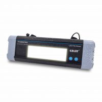
- CR System Components: Imaging plates (IP), IP readout, Monitors, Computers.
Basic CR Techniques: Image acquisition, Image quality indicators (IQIs), Display of acquired images, Optimization of displayed image, Storage of acquired and optimized image. - Digital Image Processing: Enhanced images, Signal-to-noise ratio (SNR), Artifacts and anomalies.
Image Display Characteristics: Image definition, Filtering techniques, Signal-to-noise ratio (SNR), Modulation transfer function (MTF), Grayscale adjustments, Image quality indicators (IQIs). - Image Display Characteristics: Image definition, Filtering techniques, Signal-to-noise ratio (SNR), Modulation transfer function (MTF), Grayscale adjustments, Image quality indicators (IQIs).
- Image Viewing: Image-monitor requirements, Background lighting, IQI placement, Personnel dark adaptation and visual acuity, Image identification, Location markers.
- Evaluation of CR Images: Pixel value, IQI, Artifact mitigation, System performance, Conformance to specifications, Image storage and transmission.
- Application Techniques: Multiple-view techniques (Thickness-variation parameters), Enlargement and projection, Geometric relationships (Geometric unsharpness, IQI sensitivity, Source-to-image plate distance, Focal-spot size), Localized magnification, Plate-handling techniques.
- Digital Radiography (DR) Overview: Digital radiography, Digital images (Bits/bytes, Pixels/voxels), Image file formats and compression, DR system overview, DR system capabilities (DR versus film procedural steps, Cost and environmental issues).
- DR System Components: Detector(s) used in the radiography shop (Operating procedures to use the equipment).
- Image Fidelity Indicators (System Characterization): Image quality indicators (IQIs) (hole and wire types), Line pair gages, Phantoms, Reference quality indicators (RQIs), TV test patterns.
- Detector Issues: Scatter sensitivity, Radiation exposure tolerance, Portability, Detector handling, Technique Sheets.
- Basic Digital Radiography versus Film Principles: Film versus DR images (Linearity and latitude, Contrast and resolution).
- DR System Components: X-ray and gamma-ray sources (Energy, mA, focal spot, Stability, Open and closed X- ray tubes, Filtration), Computer (Operator interface, System controller, Image processor), Monitors (CRT, LCD), Data archive (Removable media, Redundant array of inexpensive disks [RAID], Central archive).
- Image Fidelity: Measuring image fidelity (Contrast and resolution, Signal-to-noise ratio [SNR]), Image fidelity indicators (system characterization).
- Image Processing (Post-Processing): Grayscale adjustments (Windowing and leveling, Look-up tables [LUTs], Thresholding, Histogram equalization, Pseudo-color), Arithmetic, Filtering (Convolution, Low pass, High pass, Median, Unsharp mask), Region of interest (ROI).
- Detector Issues for the Detector(s) Used: Frame rate, Resolution (pixel pitch, pixel size, etc.), Blooming (bleed over), Ghosting/latent image/lag, Scatter sensitivity, Bit depth, Dynamic range and SNR, Fabrication anomalies (bad pixels, chip grades, etc.), Radiation exposure tolerance, Portability, Detector handling.
- Detector Calibrations for the Detector(s) Used: Gain and offset, Detector-specific calibration.
Monitor and Viewing Environment: Limited bit depth display, Monitor resolution, Monitor brightness and contrast, Monitor testing (Test patterns, Luminance, Contrast, DDL) Monitor calibration, Viewing area. - Monitor and Viewing Environment: Limited bit depth display, Monitor resolution, Monitor brightness and contrast, Monitor testing (Test patterns, Luminance, Contrast, DDL) Monitor calibration, Viewing area.
- Technique Development Considerations: Image unsharpness and geometric magnification (Determining required geometric magnification, Geometry and geometric unsharpness, Focal spot size measurement method, Total image unsharpness), SNR compensation for spatial resolution, Image processing.
- Detector Monitoring
- Detector Maintenance
- Use of Digital Reference Images: ASTM standards review, Use of reference images & contrast normalization.
NOTE: REGISTRATION COST IS NON-REFUNDABLE.